Research
on the effects of the ambulatory aerodynamic cervical traction apparatus
in disc patients with MRI .
Chang June, Park³ M.D. President
Airtrac MSI Inc.
#
Purpose : To evaluate the reducibility of HCD during traction,
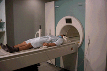
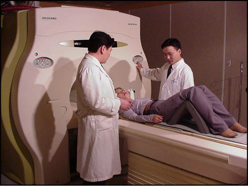
we designed an air-traction device with non-magnetic (non-metalic) materials
for use in MR machine.
# Method and Materials: Cervical
spine MR images were obtained from twenty-three paients and
seven volunteers with the application of the air-traction device
that was placed in the gantry during scanning. We designed the
air-traction device for cervical spine with an equivalent of
50-pound-weight. All of the patients and volunteers took MR
images during neutral and traction state. We compared the length
of cervical spine from C1 to C7 and examined the reducibility
of HCD on traction vs . neutral position.


# Results:
The MR images of seven volunteers showed an increasement in
the height of cervical spine from 1.5 to 4.5 mm with the
mean of 2.86mm during the traction. Seventeen of twenty-three
patients showed an increasement in height from 1 to 9mm with
the mean of 2.4mm, while remaining ten showed non response during
the traction.Three(13%) of twenty-three patients showed the
complete reduction of HCD during the traction. Ten (43%) patients
showed partially reduced HCD and remaining ten (43%) did not
show any reducibility of HCD.
# Conclusions:
The force of the air-traction
device was effective for reduction of HCD on thirteen (57%)
of twenty-three patients during the cervical spine MR imaging.
The cervical spine MRI with the application of air-traction
device can be an effective method to find reducible HCD.

* App. :
This result was not obtained in the primary or secondary
hospital but was done in University Hospital, one of the major terminal hospitals, patients from Radiology Dept. (Rehabilitation Dept., Neurosurgery dept.
Orthopedic patients, serious in-patients), so there may be difference
in actual ratio of HNP disc patients.
It
is the result with Airtrac traction treatment only, not using hyperthermal
treatment or muscle relaxants. If that therapy was done before traction,
the treatment effect may be better.
Also the MRI was taken right after the traction treatment
begun. But, if it was taken after traction treatment for 25 minutes,
the result was better. However, it was impossible because the university
hospital is too busy to delay MRI schedule for full time traction,
25 minutes.
Now, the clinical research for lumbar one is processing.
The start of lumbar research was delayed due to some technical reason.
For example, a sliding device under pelvis was needed and the MR-lumen
is not wide for patients to keep the semi-Fowler`s position.
This clinical research was reported on
11:12 AM. 29, Nov. on , in Chicago convention center. |


